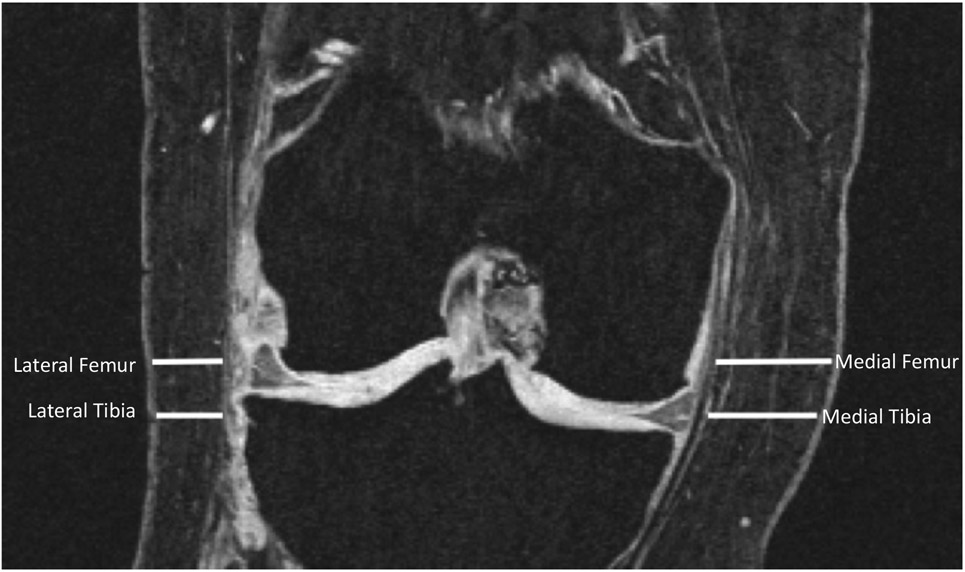
Figure 2:
A coronal reformation of the dual echo steady state (DESS) sequence. Subcutaneous fat (SCF) measurements are shown at the medial femur, medial tibia and lateral femur and lateral tibia. The tip of the medial tibial spine is used to define the axial slice level, on which medial and lateral measurements are taken.

