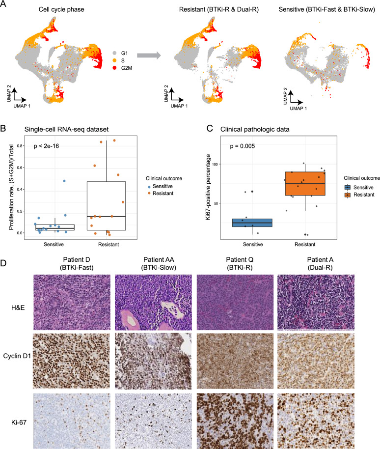
Fig. 3.
Resistant tumor cells acquire elevated proliferation rates. A Left: UMAP visualization of B cells colored by inferred cell cycle stages (G1, S, G2/M). Right: UMAP visualizations of B cells divided by clinical outcome: sensitive (BTKi-Fast and BTKi-Slow) and resistant (BTKi-R and Dual-R). Each dot represents one cell. Gray, orange, and red represent G1, S, and G2M cell cycle stages, respectively. B Boxplot shows inferred proliferation rates (y-axis) across clinical outcomes (x-axis) in single-cell RNA-seq dataset. Each dot represents one sample and is colored by clinical outcome. P-value was calculated using a generalized binomial model. C Boxplot shows proliferation rates as indicated by Ki-67-positive immunohistochemical staining across clinical outcomes from clinical pathologic data. Each dot represents one patient and is colored by clinical outcome. D Representative bone marrow images stained with hematoxylin and eosin (upper panels) or immunohistochemically stained for cyclin D1 (middle panels) or Ki-67 (bottom panels) on samples from representative patients D (BTKi-Fast), AA (BTKi-Slow), Q (BTKi-R), and A (Dual-R)

