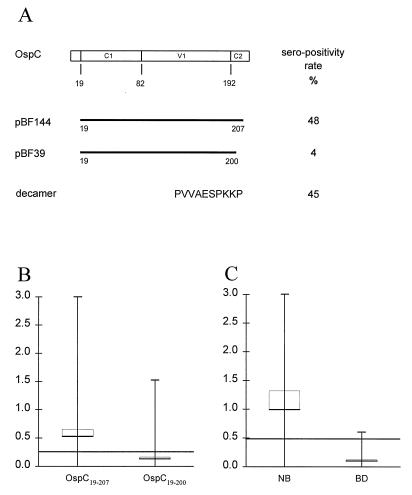
FIG. 1.
Identification of the major epitope of OspC. (A) A schematic representation of the two recombinant OspC proteins encoded by plasmids pBF144 and pBF39. Below is indicated the sequence (in single-letter code) of a synthetic peptide comprising the C-terminal 10 amino acids. The open box indicates the amino acid sequence of OspC from B. garinii DK6. C1 and C2 indicate relatively conserved regions, and V1 indicate the relatively variable region (22, 40). The NH2-terminal 19 amino acids constitute the signal peptide. Numbers below the box indicate amino acid residues. To the right is listed the percentage of sera from NB patients with a response greater than the 98% percentile for the 100 Danish blood donors. (B and C) Mean (horizontal bars) and range (vertical bars) of the IgM reactivity against OspC19–207 and OspC19–200 of the 100 serum samples from NB patients (B) and the anti-PVVAESPKKP IgM reactivity of sera from the 100 NB patients and from Danish blood donors (BD) (C); the thin brackets represent the 95% confidence interval. The 98% specific cutoff levels based on the examination of 100 Danish blood donors were optical densities of 0.230 for anti-OspC19–207 and anti-OspC19–200 IgM reactivity and 0.450 for anti-peptide IgM reactivity.

