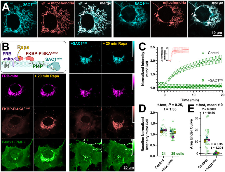
Figure 2.
An engineered mitochondria-targeted SAC1 is active in cells. (A) SAC1mito co-localizes with the mitochondrial marker, CAX8AN29×2-mCherry (pink). Images show representative equatorial confocal sections of two COS-7 cells. (B) Mitochondrial PI4P synthesis was induced by the addition of rapamycin, which induces dimerization of over-expressed mCherry-FKBP-PI4KC1001 with iRFP-FRB-mito; this initiates ectopic PI4P synthesis on mitochondria outer membranes, detected with EGFP-P4Mx1 biosensor. The accumulation is blocked by co-expression of tagBFP2-SAC1mito. Images show equatorial confocal sections of COS-7 cells before or after addition of rapamycin. P4Mx1 intensity is normalized to the average cell intensity, as quantified in B. (C) PI4P accumulation on mitochondria is blocked by SAC1mito, despite equally efficient PI4KAC1001 recruitment (inset). (D) The slightly decreased baseline level of PI4P at mitochondria is not statistically significant by nested t-test. Smaller green points show individual cells (7–11 cells/experiment) and larger colored-shapes show means of each of four experiments. Grand means ± SEM are also indicated. (E) Complete reduction of mitochondrial PI4P synthesis, demonstrated by a grand mean of the area under the curve for SAC1mito-expressing cells of 0.24 ± 0.20 that is not significantly different from 0 by t-test.

