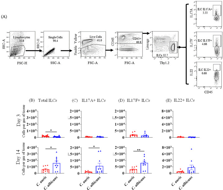
Fig 4.
Absolute number of ILCs in the skin of C. auris- and C. albicans-infected mice groups. Mice were infected intradermally with C. auris 0387 or C. albicans SC5314. Mice were euthanized after 3 and 14 days post-infection to determine ILCs. (A) Flow gating strategy for ILCs is shown here. Absolute numbers of (B) total ILCs, (C) IL-17A+ ILCs, (D) IL-17F+ ILCs, and (E) IL-22+ ILCs in C. auris- or C. albicans-infected mice skin tissue after 3 and 14 days post-infection are shown. Ten mice were used for each group and data represented were mean ± standard error of the mean for each group. Statistical significance was calculated using Mann-Whitney U test. *P ≤ 0.05, **P ≤ 0.01 were considered as significant. ILC, innate lymphoid cell.

