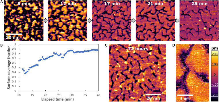
Fig. 1. In situ AFM images of gibbsite nucleation and growth at the mica-water interface.
(A) In situ AFM image sequence from movie S1, showing the growth and coalescence of clusters to create an extended film, with a persistent network of gaps, obtained in a pH 3.7 solution (1 mM AlCl3 + 0.2 mM HCl) at 65°C. (B) Corresponding plot of surface coverage versus elapsed time (relative to time when temperature began rising to 65°C). (C) Image of a film aged ex situ at the same conditions for 72 hours and then imaged in situ at room temperature. (D) High-resolution in situ AFM of a sample aged at similar conditions for several days, revealing atomistic details of the surface crystallinity and morphology of the gibbsite monolayer, including extensive crystalline defects. Image obtained at room temperature, after growth.

