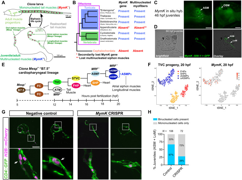
Fig. 2. MymK is required for multinucleation of postmetamorphic muscles in the tunicate Ciona.
(A) Diagram of biphasic life cycle of ascidians (sessile tunicates) like Ciona. The motile larvae have strictly mononucleated tail muscles during the dispersal phase. After settlement and metamorphosis, tail muscle cells undergo programmed cell death and are reabsorbed, while dedicated muscle progenitors set aside in the larva differentiate to form the multinucleated siphon and body wall muscles of the juvenile. Muscles surrounding and emanating from the oral and atrial siphons are derived from distinct cell lineages in the larva. Only those from the atrial siphon are derived from the Mesp+ B7.5 lineage [in (E)]. (B) Cladogram of extant chordates showing correlation between the presence of MymK gene and muscle multinucleation in different clades. (C) Whole-mount mRNA in situ hybridization showing MymK expression in developing atrial siphon muscle (ASM) and oral siphon muscle (OSM) cells in metamorphosing juveniles. Smaller arrows indicate autofluorescent tunic cells. (D) C. robusta juvenile developed from a zygote transfected with a MymK promoter reporter plasmid, labeling ASMs and longitudinal body wall muscles (LoM). (E) Diagram of the B7.5 lineage in C. robusta, based on conclusions from (18). FC, founder cell; TVC, trunk ventral cell; ATM, anterior tail muscle cell; STVC, secondary TVC; FHP, first heart precursor; SHP, second heart precursor; ASMF, atrial siphon muscle founder cell; ASMP, atrial siphon muscle precursor; oASMP, outer ASMP; iASMP, inner ASMP. Asterisk indicates that both FCs give rise to identical lineages. MRF, myogenic regulatory factor (MyoD ortholog). (F) t-distributed stochastic neighbor embedding (tSNE) plots based on information from (16) showing MymK expression mapped onto TVC progeny clusters at 20 hpf. MymK is expressed exclusively in ASMPs and especially enriched in outer ASMPs. Abbreviations same in (E). (G) Representative Z-projection confocal fluorescence images of 84 hpf negative control (transfected with Mesp > Cas9 only, no sgRNAs) juveniles alongside same-age juveniles in which MymK was targeted for mutagenesis specifically in the B7.5 lineage. MymK CRISPR: zygotes transfected with Mesp > Cas9 and U6 > MymK-sgRNA vectors. Muscle plasma membranes and nuclei labeled by MRF > CD4::GFP and MRF > H2B::mCherry, respectively. Arrows in negative control panels showing development of typical binucleated myofibers that is inhibited upon MymK CRISPR. (H) Data from scoring of juveniles represented in (G) showing reduced frequency of binucleated atrial siphon/longitudinal myofibers in MymK CRISPR juveniles. N, numbers of juveniles assayed for each condition. Scale bars, 50 μm.

