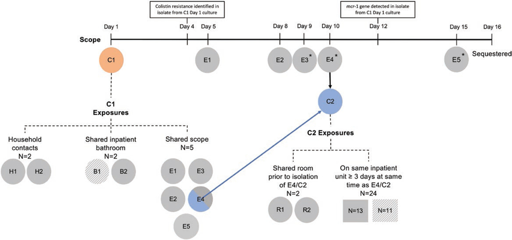
Figure 1.
Exposure investigation timeline and surveillance results. Patients exposed to the duodenoscope are shown below the timeline, from duodenoscope use during case 1’s (C1) procedure on day 1 through sequestration of the scope on day 16. The following designations were used: C for case-patients; E denoting endoscopic retrograde cholangiopancreatography (ERCP) exposure; R denoting roommate exposure; B indicating bathroom exposure, and N indicating patients identified as unit contacts. An asterisk indicates those patients who either had a stent placed during the ERCP (E3 and E4/C2) or who had an indwelling stent that was left in place following the ERCP (E5). Exposed patients who were identified as cases of mcr-1 are shown in color (orange for case 1 [C1], blue for case 2 [E4/C2]). Secondary exposures to C1 and E4/C2 were identified using a definition of shared environment informed by public health recommendations. Together, these individuals constituted the cohort for the exposure investigation. Exposed patients who were tested and had an mcr-1–negative result are shaded in solid gray. Exposed patients for whom no information was available are shown in hatched gray (eg, patient died or was discharged and lost to follow-up). Of the 7 individuals identified as healthcare-associated contacts of C1, all had been discharged from the hospital to home at the time the investigation was initiated, apart from E4/C2. Of the 26 contacts of E4/C2, 9 had been discharged to home, 13 had been transferred to a long-term acute care hospital or rehabilitation hospital, and 4 had died either in the hospital or since discharge; swabs could not be obtained from 11 of these 26 contacts.

