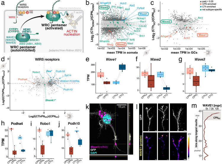
Figure 3: Core components and regulatory elements of the WAVE regulatory complex are subcellularly enriched in GCs of CPN and CThPN, suggesting local assembly and subtype-specificity of the machinery regulating actin branching.
(a) Schematic of the composition, regulation, and function of the WRC (also termed SCAR complex), which is activated upon association with WRC-interacting regulatory sequence (WIRS)-containing receptors and is important for actin nucleation and branching. Adapted from Rottner 20211. (b) WRC core genes and regulators: transcript abundances and subtype-specific expression in CPN and CThPN somata. (c) WRC core genes and regulators: transcript abundances and subtype-specific expression in CPN and CThPN GCs. (d) Localization plot showing subtype-specific localization of transcripts encoding high confidence WIRS-containing proteins. (e-g) Normalized transcript abundances of Wave1, Wave2, and Wave3, indicating their subtype-specificity and subcellular transcript localization. (h-j) Normalized transcript abundances of select WIRS-containing transcripts, showing their subtype-specificity and subcellular localization. (k) Maximum intensity projection (10 μm total z depth) of representative RNAscope confocal images of Wave1 in GFP-positive cultured CPN. Localization of the magenta Wave1-positive puncta in the tip of the neurite is confirmed by respective orthogonal z-axis views along the sides. (l) Maximum intensity projection (10 μm total z depth) of representative immunocytochemistry for GFP (top panels) and WAVE1 (bottom panels) in GCs of cultured CPN. WAVE1 protein is detected as focal puncta at the tip of CPN GCs. (m) Quantification of WAVE1 immunocytochemical signal intensity (mean grey value, mgv ± sem, n=11) in the longest neurite of cultured CPN (n = 11), from proximal (bottom) to distal (top) end of the neurite. Grey box indicates localization of GC as assessed by parallel immunolabeling for actin.

