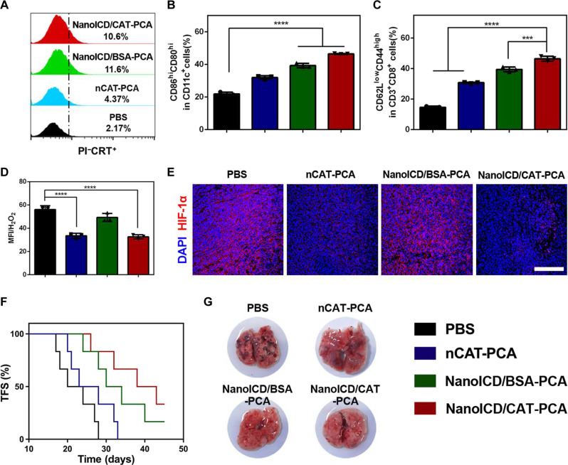
Fig. 7. NanoICD/CAT-PCA activates ICD and remodels immunosuppressive TME in vivo.
(A) Pre-apoptotic CRT exposure on tumors from mice treated with PBS, nCAT-PCA, NanoICD/BSA-PCA, and NanoICD/CAT-PCA. (B) The population of CD80+CD86+ DC cells (gated on CD11c+) in tumor-draining lymph nodes of mice after receiving different treatments. (C) The population of CD44+CD62L− effector memory cells (gated on CD3+CD8+) in spleen of mice after receiving different treatments. (D) Flow cytometric analysis showing H2O2 concentrations in tumor tissues after different treatments. (E) Immunofluorescence images showing HIF-1α concentrations in tumor tissues after different treatments. (F) The TFS of 4T1-bearing BALB/c mice after different treatments as indicated. (G) The lung metastases in mice after the indicated treatments. Scale bar, 200 μm. Data are presented as means ± SD from three independent experiments for (B) to (D) (biological replicates, n = 3) and six independent experiments for (F) (biological replicates, n = 6). Significant levels are ***P < 0.001 and ****P < 0.0001. Scale bar, 200 μm.

