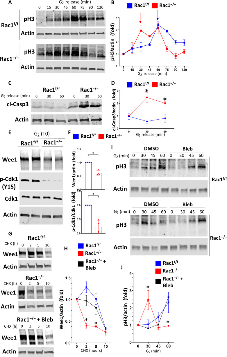
Fig. 9. Rac1 via actomyosin regulates the G2-M checkpoint kinase Wee1 to prevent premature mitotic entry.
(A and B) CD cells were G2-synchronized using RO-3306, and lysates were collected at the indicated time points after RO-3306 washout (“G2 release”). Lysates were immunoblotted for pH3 to monitor mitotic entry. Three repeat experiments were quantified in (B). Fold change values ± SD. Arrows highlight the first pH3 peak of the respective groups indicating mitotic entry. (C and D) G2-synchronized CD cells were immunoblotted for cleaved caspase 3 (cl-Casp3) after G2 release and quantified in (D) as fold change values ± SD (n = 3). Asterisk (*) denotes between-group significance at the corresponding time point. (E and F) CD cells were G2-synchronized, and lysates were obtained immediately upon G2 washout (G2, T0) and immunoblotted in biological duplicates. p-Cdk1 Y15: phosphorylated Cdk1 tyrosine-15. Three repeat experiments were quantified in (F). Fold change values ± SD. (G and H) Asynchronous Rac1f/f (+DMSO) and Rac1−/− [+DMSO or blebbistatin (5 μM)] CD cells were treated with cycloheximide (CHX; 100 μg/ml), and lysates were obtained at the indicated time points and immunoblotted for Wee1. Three repeat experiments are quantified in (H) as fold change ± SD. Asterisk (*) denotes significance between Rac1−/− and Rac1f/f or blebbistatin-treated Rac1−/− at the corresponding time point. (I and J) Rac1f/f and Rac1−/− CD cells were G2-synchronized and treated with vehicle (DMSO) or 5 μM blebbistatin upon G2 release. Lysates were collected at the indicated time points and immunoblotted for pH3 to monitor mitotic entry. Three independent repeat experiments are quantified (for Rac1f/f, only the vehicle control is shown) in (J) as fold change values ± SD. Asterisk (*) denotes significance between Rac1−/− and Rac1f/f or blebbistatin-treated Rac1−/− at the corresponding time point. *P < 0.05.

