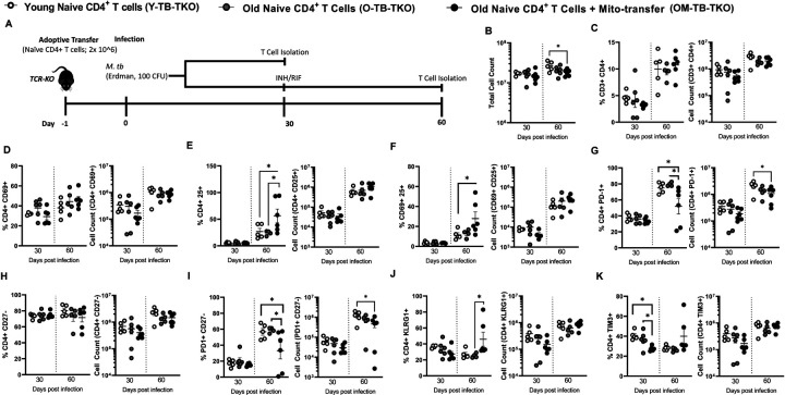
Fig. 3. Mito-transfer in naïve CD4 T cells from old mice protects mice against pathogens & promotes protective T cell phenotypes in the lung.
A) 2.0 × 10⁶ naïve CD4+ T cells from young mice and old mice with or without mito-transfer, were tail vein injected in TCR-KO mice, that were the subsequently infected M.tb. A subset of mice were euthanized at 30 days post-infection. Another subset of mice were treated with INH/RIF for an additional 30 days (up to 60 days post-infection). The absolute and percentage of various CD4+T cell subsets in the lung of M.tb infected mice were evaluated at both time points (30 and 60 days post-infection). B) Absolute cell count of the digested lung from TCR-KO mice infected with M.tb. The percents and absolute counts of C) CD3+CD4+ T cells, D) CD4+CD69+,E) CD4+CD25+, F) CD4+ CD69+ CD25+, G) CD4+ PD-1+, H) CD4+ CD27−, I) CD4+ PD-1+ CD27−, J) CD4+ KLRG1+, K) CD4+ TIM3+ subsets isolated from TCR-KO mice infected with M.tb, at 30 and 60 days post-infection. p< 0.05 = significant (*) using one-way-ANOVA.

