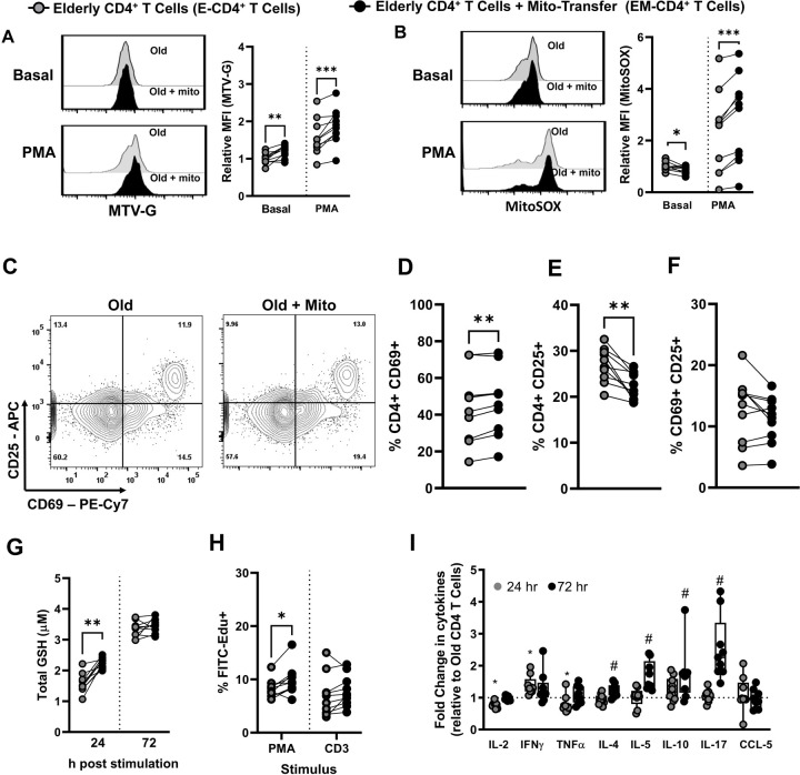
Fig. 5. T cell activation and proliferation improved in CD4+ T cells from old humans after mito-transfer.
Donor mitochondria isolated from primary human neonatal dermal fibroblasts were transplanted into CD4+ T cells from elderly humans. Flow cytometry histograms and the relative fold changes of A) MTV-G, B) MitoSOX stained, unstimulated, or PMA/Ionomycin stimulated human CD4+ T cells with or without mito-transfer. C) Contour plots of CD25 v CD69 expression on CD4+ T cells with or without mito-transfer, after PMA/Ionomycin stimulation (24h). The percentages of D) CD69+, E) CD25+, and F) CD69+CD25+ human CD4+ T cells after PMA/Ionomycin stimulation (24h). G) Total GSH produced by CD4+ T cells with or without mito-transfer, stimulation with PMA/ Ionomycin, at 24h and 72 h. H) The number of EdU+ cells in PMA/Ionomycin-stimulated human CD4+ T cell cultures with or without mito-transfer. I) The relative fold change in cytokine production of human CD4+ T cells with or without mito-transfer, after stimulation with PMA/Ionomycin for 24 h and 72 h. 9–10 biological replicates per group with p ≤ 0.05 = *, p ≤ 0.01 = **, or p ≤ 0.001 = *** using paired Student’s t-test.

