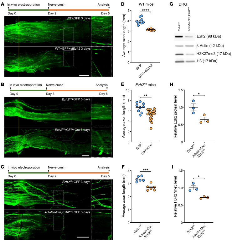
Figure 2. Upregulation of Ezh2 contributes to spontaneous axon regeneration of DRG neurons in vivo.
(A–C) Top: Experimental timeline. Bottom: Representative images of sciatic nerves showing that Ezh2 knockdown (A) or knockout (B) in L4/5 DRGs or sensory neuron-specific knockout of Ezh2 (C) impairs spontaneous axon regeneration of DRG neurons in vivo. The right column displays enlarged images of the areas in white, dashed boxes on the left. The crush sites are aligned with the yellow line. Scale bars: 1 mm, 0.5 mm for enlarged images. (D) Quantification of lengths of regenerating axons in A (unpaired, 2-tailed t test; P 0.0001; n = 9 mice for control, n = 10 mice for Ezh2 knockdown). (E) Quantification of lengths of regenerating axons in B (unpaired 2-tailed t test; P = 0.0011; n = 9 mice for control, n = 18 mice for Ezh2 knockout). (F) Quantification of lengths of regenerating axons in C (unpaired 2-tailed t test; P = 0.0003; n = 6 mice for both). (G) Representative immunoblotting showing successful knockout of Ezh2 and downregulation of H3K27me3 in DRG neurons of Advillin-Cre;Ezh2fl/fl mice. (H) Quantification of relative protein levels of Ezh2 in G (unpaired 2-tailed t test P = 0.0436; n = 3 independent experiments). (I) Quantification of relative levels of H3K27me3 in G (unpaired 2-tailed t test; P = 0.0137; n = 3 independent experiments). siEzh2, siRNAs targeting Ezh2 mRNA. *P 0.05, **P 0.01, ***P 0.001, ****P 0.0001.

