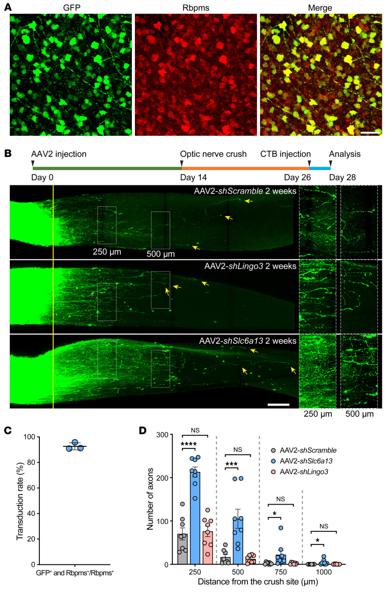
Figure 7. Slc6a13 loss-of-function promotes optic nerve regeneration.
(A) Representative immunofluorescence of whole-mount retinas showing high transduction efficiency of AAV2-shSlc6a13-EGFP in RGCs by intravitreal injection. Whole-mount retinas were stained with anti-GFP (green) and anti-Rbpms (red). Scale bar: 50 μm. (B) Top: Experimental timeline. Bottom: Representative images of optic nerves showing that knockdown of Slc6a13, but not Lingo3, modestly promotes optic nerve regeneration 2 weeks after optic nerve crush. Columns on the right display enlarged images of the areas in the white, dashed boxes on the left, showing axons at 250 and 500 μm distal to the crush sites, which are aligned with the yellow line. Yellow arrows indicate longest axons in each nerve. Scale bars: 100 μm, 50 μm for enlarged images. (C) Quantification of the percentage of GFP-positive RGCs in A. The average transduction rate was 92.65% ± 2.743% (n = 3 mice; 8 fields were analyzed for each mouse). Data represent mean ± SD. (D) Quantification of optic nerve regeneration in B (1-way ANOVA followed by Tukey’s multiple comparisons; P 0.0001 at 250 and 500 μm, P = 0.0248 at 750 μm, P = 0.0263 at 1,000 μm; n = 8 mice for all). *P 0.05, ***P 0.001, ****P 0.0001.

