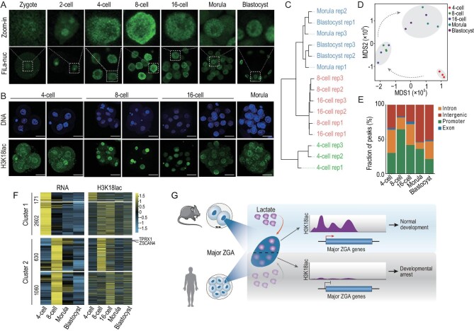
Figure 6.
H3K18lac-enriched ZGA markers in human 8-cell embryos. (A) Fluorescence images of human embryos from different stages expressing FiLa in nuclei. (B) IF of H3K18lac of human 4-cell, 8-cell, 16-cell, and morula embryos. Scale bars, 50 μm. (C) Unsupervised clustering of H3K18lac signals among the embryos at the stages indicated. (D) MDS analysis of samples in the indicated stages. Different colors indicate different developmental stages. (E) Ratio of fraction of the human genome covered by H3K18lac reads in the indicated developmental stages, and percentages of H3K18lac peaks assigned to the promoter, intron, exon, and intergenic regions. (F) Heatmaps of H3K18lac enrichment (right panel) in 8-cell signature genes indicated in human embryos; left panel, corresponding RNA levels. (G) Lactate in nuclei when major ZGA occurs in humans and mice during preimplantation development and a model of lactate-derived H3K18lac regulating major ZGA.

