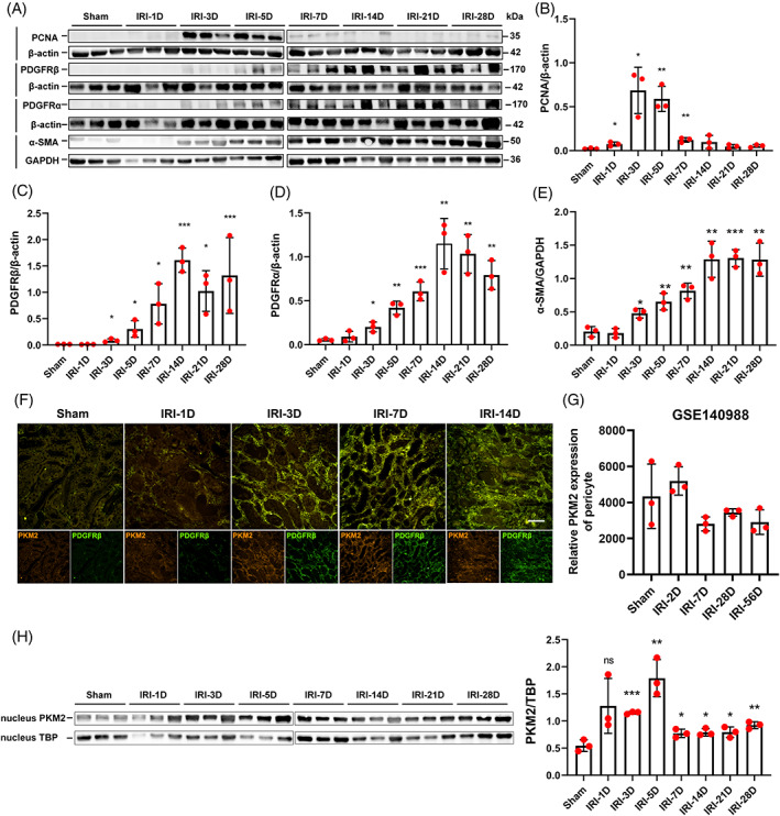
FIGURE 2.

Pericytes continually expressed PKM2 during the progression of acute kidney injury‐chronic kidney disease. (A‐E) Expression of PCNA, PDGFRβ, PDGFRα, and α‐SMA in IRI kidney samples, as detected by Western blotting (n = 3). (F) The location of PDGFRβ and PKM2 expression in PDGFRβ‐iCreERT2; mTmG mice IRI kidney samples, as detected by immunofluorescence staining. (G) PKM2 mRNA was continually expressed in mouse renal pericytes after IRI (dataset GSE140988) (n = 3). (H) The expression of PKM2 in the nucleus of renal PDGFRβ‐positive pericytes sorted from IRI kidney samples, as detected by Western blotting (n = 3). The data are presented as follows: Error bars, mean ± SD; Scale bar = 100 μm; ns, not significant; *p < 0.05, **p < 0.01, ***p < 0.001 versus Sham. GAPDH, glyceraldehyde‐3‐phosphate dehydrogenase; IRI, renal ischaemia–reperfusion injury; PCNA, proliferative cell nuclear antigen; PDGFRβ, platelet‐derived growth factor receptor β; PKM2, pyruvate kinase M2; TBP, TATA binding protein.; α‐SMA, alpha smooth muscle actin.
