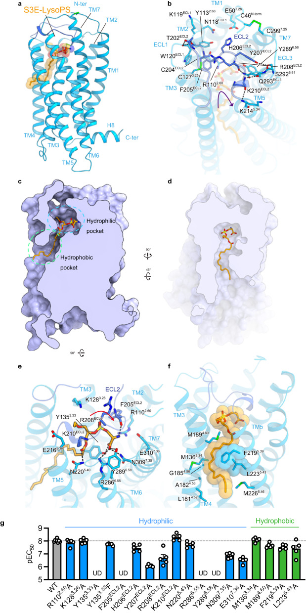
Fig. 2. S3E-LysoPS binding mode.
a Overall structure of the S3E-LysoPS-bound receptor. Disulfide bonds are shown as sticks. b Interactions between extracellular loop (ECL)2 and transmembrane helices (TMs). Black dashed lines indicate hydrogen-bonding interactions. c, d Cross-sectional views of the ligand-binding pocket, viewed from the membrane plane (c) and the extracellular side (d). e, f Binding mode of S3E-LysoPS in the hydrophilic (e) and hydrophobic (f) pockets of the receptor. g Mutagenesis data for identifying the S3E-LysoPS interaction residues (n = 8 for the wild type and n = 5 for the mutants). Values are shown as the mean ± s.e.m. from at least three independent experiments performed in triplicate. Source data are provided as a Source Data file.

