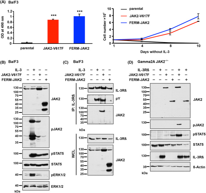
Fig. 2.

FERM‐JAK2 transforms Ba/F3 cells and activates STAT5 via a non‐canonical pathway. (A) Left panel: Proliferation of parental Ba/F3 cells and Ba/F3 cells expressing FERM‐JAK2 or JAK2‐V617F in the absence of IL‐3 was quantified by the relative optical density (OD) after 96 h using an MTS (3‐(4,5‐dimethylthiazol‐2‐yl)‐2,5‐diphenyltetrazolium bromide)‐based assay. Right panel: Absolute cell numbers over time were measured in the absence of IL‐3 by trypan blue exclusion (n = 3). ***P < 0.001 compared to parental cells by Student's t test. Data are shown as mean ± standard deviation (SD). (B) Immunoblot analysis of serum‐starved Ba/F3 cells expressing FERM‐JAK2 or JAK2‐V617F. A representative image of n = 2 two independent experiments is shown. (C) IL‐3Rβ immunoprecipitation (IP) analysis of Ba/F3 cells expressing FERM‐JAK2 or JAK2‐V617F. pY, phosphotyrosine; WCL, whole cell lysate. A representative image of n = 3 three independent experiments is shown. (D) Immunoblot analysis of Gamma2A cells stably expressing mock vector, FERM‐JAK2 or JAK2‐V617F in combination with or without IL‐3Rβ chain. A representative image of n = 3 three independent experiments is shown.
