Figure 1.
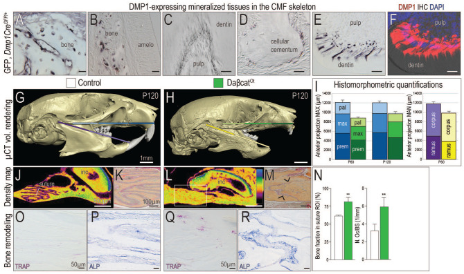
Maxillary and mandibular hyperostosis result from constitutive activation of Wnt/β-catenin signaling in DMP1-expressing cells. Using Dmp1CreGFP/+ mice, immunostaining for green fluorescent protein (GFP) was performed in mineralized tissues of the craniomaxillofacial (CMF) skeleton, including (A, B) alveolar bone, (C) odontoblasts, and (D) cementoblasts near the tooth root apices. In near-adjacent sections, the patterns of (E) GFP immunostaining and (F) Dmp1 immunostaining are indistinguishable. Micro–computed tomography (µCT) imaging of postnatal day 120 (P120) (G) control and (H) daβcatOt mice. Colored bars correspond to (I) histomorphometric quantifications of the premaxilla, maxilla, palatine bones in the maxilla, and the corpus and ramus in the mandible. Density mapping of µCT sagittal sections through (J) control and (L) daβcatOt maxillae, where the thickest bone is blue and the thinnest portion is purple (see scale). Pentachrome staining of representative tissue sections through (K) control and (M) daβcatOt premaxillary-maxillary sutures. (N) Quantification of a region of interest (ROI) in the suture, where the percentage of bone was calculated and quantification of Tartrate-resistant acid phosphatase positive (TRAP+) osteoclasts as a function of the perimeter of bone surface. (O) TRAP staining and (P) Alkaline phosphatase (ALP) activity shown in representative tissue sections through the premaxillary suture of controls and (Q, R) and daβcatOt mice. amelo, ameloblasts; mx, maxilla; pal, palatine; po, periosteum; premx, premaxilla. Scale bars: as indicated. **P < 0.01.

