Figure 4.
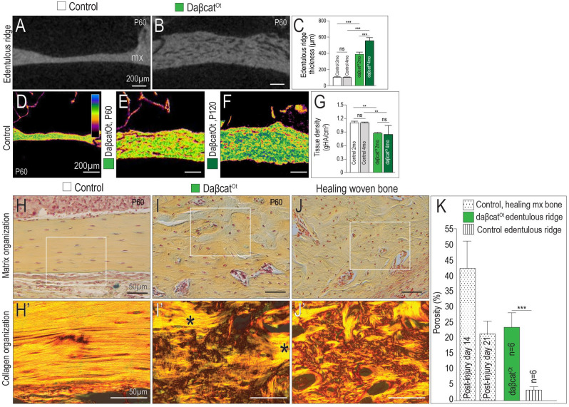
Constitutively activated Wnt/β-catenin signaling transforms lamellar into woven bone. Representative sagittal micro–computed tomography (µCT) sections through the maxillary edentulous ridge of (A) control and (B) daβcatOt mice. (C) Quantification of edentulous ridge thickness. Density mapping of µCT sagittal sections through (D) control (P60), (E) daβcatOt mice at postnatal day 60 (P60), and (F) daβcatOt mice at postnatal day 120 (P120). (G) Quantification of tissue density as determined by µCT analysis. Pentachrome staining and picrosirius red staining of representative sagittal tissue sections, viewed under polarized light to visualize collagen fiber orientation in the maxillary edentulous ridges of (H, H′) controls, (I, I′) daβcatOt mice (asterisks indicate islands of lamellar bone surrounded by woven bone), and (J, J′) healing maxillary bone from a wild-type mouse. (K) Quantification of porosity in bone as a function of injury and genotype. ns, not significant. **P< 0.01. ***P < 0.001.

