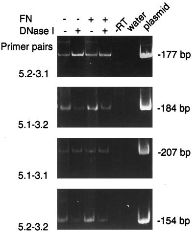
FIG. 1.
Expression of hBD-1 mRNA in cultured HGE cells stimulated with F. nucleatum cell wall extract (FN) as analyzed by 35 cycles of RT-PCR. HGE cells were cultured in serum-free keratinocyte growth medium and incubated overnight with 100-μg/ml F. nucleatum cell wall extract (+) or were unstimulated (−). DNase I was used in some samples to digest the genomic DNA possibly contaminating the RNA samples. PCR products, as described in Materials and Methods, were separated by electrophoresis on a 1.5% agarose gel and stained with ethidium bromide. For the locations of the four primers, see Fig. 2. −RT denotes a control in which the reverse transcriptase enzyme was omitted. The water lane was the negative control. The molecular sizes of PCR products from RNA samples and the hBD-1 plasmid (plasmid lane) were predicted from the sequences and were in accordance with the molecular size markers (φX174RF HaeIII fragments [not shown]).

