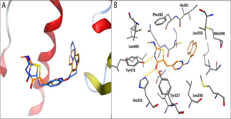
Figure 12.
Cartoon representation of docking of TZD # 14 onto the PPAR-γ binding site of the PDB: 1ZGY crystal structure. (A) shows TZD # 14 (orange sticks) aligned with the cocrystallized ligand (blue sticks). (B) shows the binding modes. Hydrogen bonding is shown as orange dotted lines.

