
Figure 1. Ipsilateral boost of SARS-CoV-2 mRNA vaccine induces more RBD-specific GCBCs.
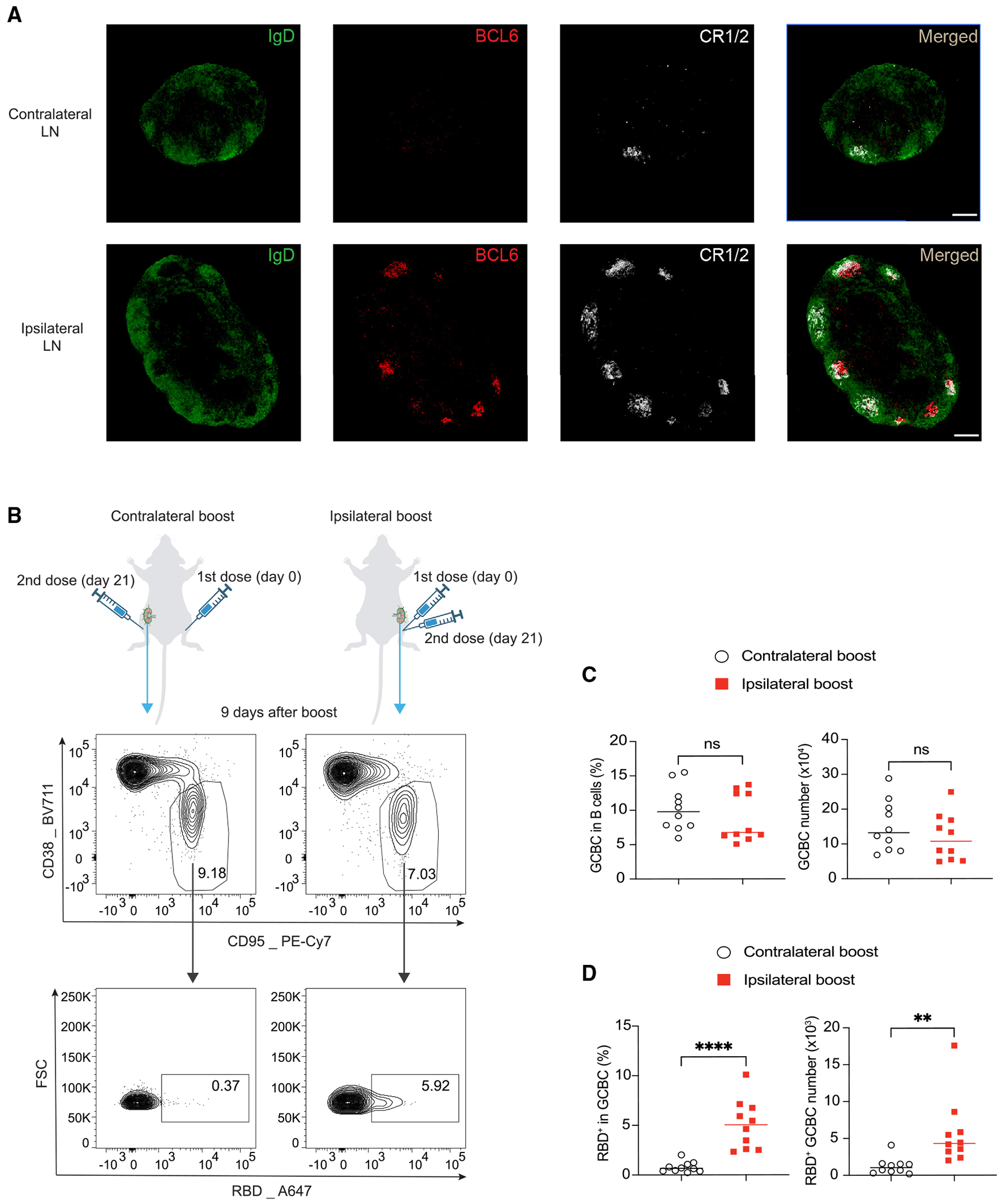
(A) Immunofluorescence staining of ipsilateral dLNs and contralateral LNs 3 weeks after a single dose of mRNA vaccine immunization. Shown are popliteal LNs. Scale bar represents 200 μM. n = 3 from two independent experiments.
(B–D) Mice were vaccinated with 0.2 μg SARS-CoV-2 mRNA vaccine and boosted with the same dose of mRNA vaccine 21 days later in the same limb (ipsilateral boost) or the opposite limb (contralateral boost). 9 days after booster vaccination, total GCBCs and SARS-CoV-2 RBD-specific GCBCs in the dLNs to the booster were analyzed by flow cytometry. Representative flow cytometry plots (B) and statistical analysis (C and D) are shown. n = 10 from three independent experiments. Each dot represents one mouse, with lines depicting the median. Data were analyzed by unpaired t test. **p < 0.01; ****p < 0.0001; ns: not significant.