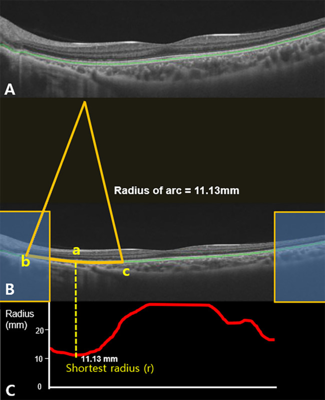
Fig. 1.

The prototype software to automatically measure radii of arcs on retinal pigment epithelium (RPE) segmentation line on OCT images. (A) The machine software automatically detects the RPE segmentation line (green line). (B) Given that the raw OCT images were elongated to twice the size in the vertical dimension in the machine software, the horizontal to vertical pixel ratio was corrected to 1:1. The region of interest was defined by excluding 100 pixels in the horizontal dimension on both sides of the image (Yellow box). The arc at the pixel (a) was made by connecting two pixels (b and c) apart by 1.4 mm [100 pixels apart from the pixel (a)] on RPE segmentation line. (C) The radius of arc was calculated at every pixel on the RPE segmentation line in the region of interest and they were presented as the graph (red line). Among graphs of the radii of arc (red line), the shortest radius was named ‘r’ (mm) (Fig. 1C). The average ‘r’ of the 12 radial OCT scans was designated as ‘R’ (mm)
