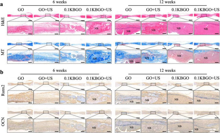
Fig. 6.
Histological analysis of newly formed bone tissue at 6 weeks and 12 weeks after surgery. (a) H&E and MT staining of demineralized calvarial sections. Higher magnification images are taken from the areas enclosed by a square in the upper row. (b) Runx2 and OCN immunohistochemical staining of demineralized calvarial sections. Higher magnification images are taken from the areas enclosed by a square in the upper row. WB: woven bone, NB: newly formed bone. The white arrow represents newly formed bone marrow. Scale bars represent 1 mm (lower magnification) and 200 μm (higher magnification)

