Figure 5.
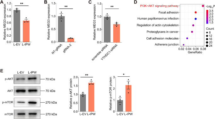
IPW inhibits MEG3 to activate the AKT/mTOR pathway in RPE cells. (A) MEG3 expression detected by qPCR in fRPE cells transduced with L-IPW compared with cells transduced with L-EV (n = 3 per group). (B) MEG3 expression detected by qPCR in fRPE cells cotransfected with the dCas13b-ALKBH5 plasmid and NT-gRNA/gRNA-2 (n = 3 per group). (C) MEG3 expression detected by qPCR in fRPE cells transfected with scramble siRNA or YTHDC1-siRNA (n = 3 per group). (D) Kyoto Encyclopedia of Genes and Genomes (KEGG) plot of enriched pathways in fRPE cells transduced with L-IPW compared with cells transduced with L-EV. (E) Immunoblotting of p-AKT, AKT, p-mTOR, and mTOR in fRPE cells transduced with L-EV or L-IPW (n = 3 per group). Data are presented as mean ± SEM. Two-tailed Student t test was used. * P < 0.05 and ** P < 0.01.

