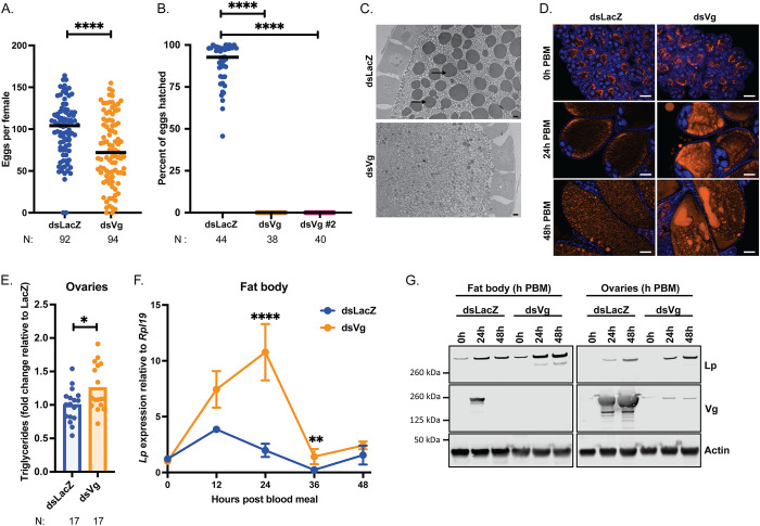
Fig 2. Vg knockdown upregulates Lp expression and enhances lipid deposition into oocytes.
(A) Following Vg knockdown females develop fewer eggs, as identified by dissection of ovaries of virgin females at 3–7 days PBM; each dot represents eggs per female; N = number of females, pooled from five biological replicates (Mann-Whitney: **** = p < 0.0001). (B) Vg knockdown causes complete infertility of mated females; Vg was targeted by two different dsRNAs; each dot represents percent hatch rate per female; N = number of females, pooled from three biological replicates (Kruskal-Wallis with Dunn’s multiple comparisons test: **** = p < 0.0001). (C) Transmission electron microscopy of developing oocytes showing a lack of yolk granules (arrows) upon Vg knockdown at 24h post blood meal; scale bar = 2 μm (representative images, two biological replicates). (D) Fluorescent microscopy showing an accumulation of neutral lipids (LD540, orange) in developing oocytes upon Vg knockdown; DNA (DAPI) in blue; scale bar = 50 μm (representative images, three biological replicates). (E) Triglyceride levels measured in dsLacZ and dsVg ovaries at 48h post blood meal and normalized to mean dsLacZ levels in each replicate; each dot is representative of ovaries from three females; N = number of samples of three tissues, pooled from three biological replicates (Unpaired t-test: * = p < 0.05). (F-G) Vg knockdown results in an increase in Lp levels in the fat body at the mRNA (samples of ten tissues, four biological replicates (REML variance component analysis: ** = p < 0.01; **** = p < 0.0001)) (F) and protein (samples of five tissues, representative blots, three biological replicates) (G) levels in the fat body and ovaries.

