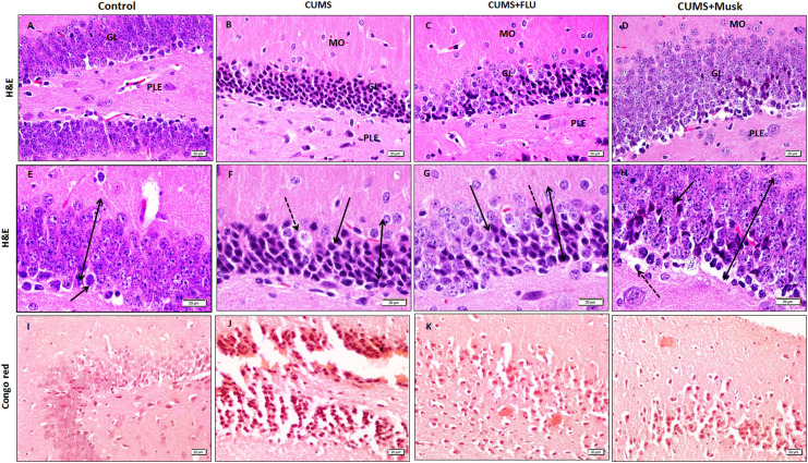
Figure 6.
The hippocampal DG is formed of the PLE, the GL, and the MO. The thickness of the GL layer appears smaller in the CUMS compared to the control (bi-head arrow). Some cells appear vacuolated cells (interrupted arrow) are observed. Note the increased number of the immature cell that have darkly stained nuclei (arrow). Amyloid deposition with salmon red coloration observed as patches as well as aggregation around the granular cell of the DG (A-D, I-L x400, E-H x1000). CUMS indicates chronic unpredictable mild stress, DG, dentate gyrus; FLU, fluoxetine; GL, granular cell layer; M; musk; MO, molecular; PLE, pleomorphic layer.

