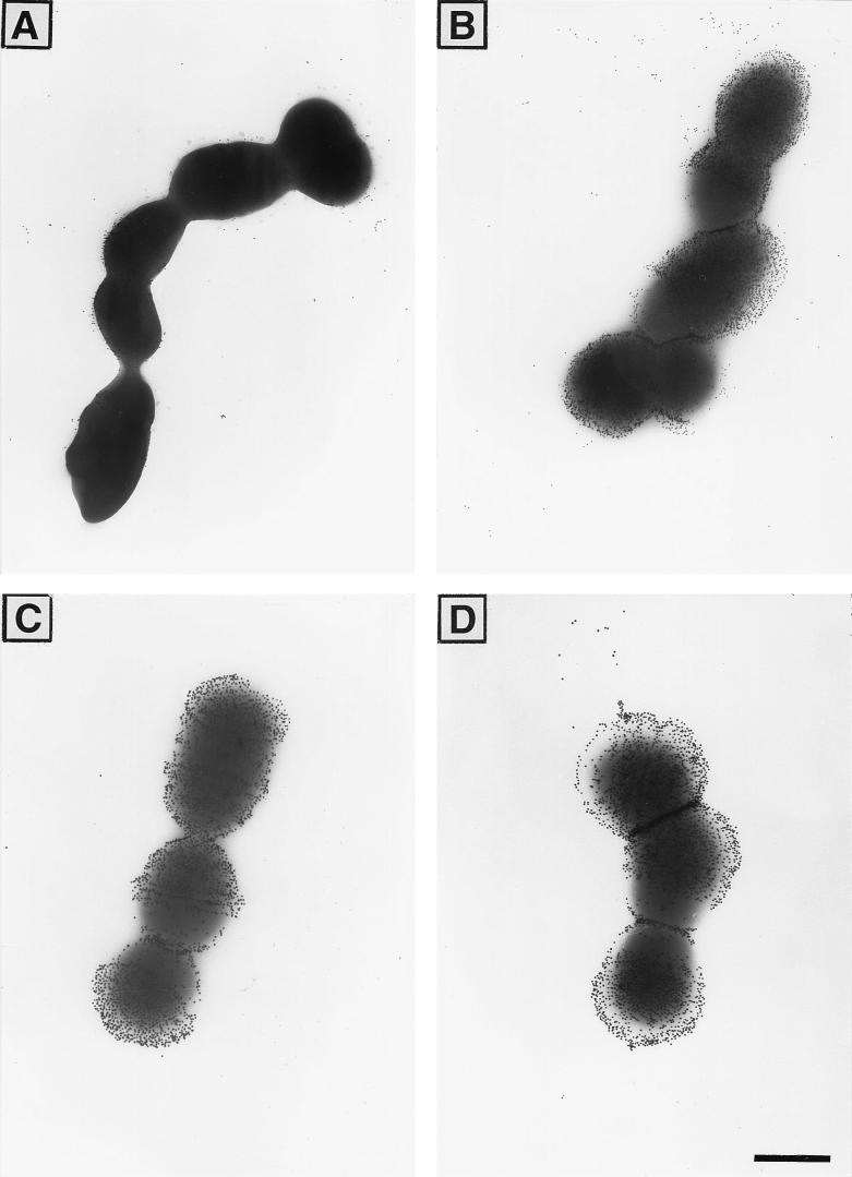
FIG. 3.
EM photographs of GBS with one-repeat alpha C protein (A and C) and nine-repeat alpha C protein (B and D) at the cell surface, incubated with one-repeat alpha C protein-specific rabbit antiserum (A and B) or with rabbit antiserum to CPS type Ia (C and D). For protein staining, 15-nm-diameter-gold-labelled protein A was used, and for the CPS type Ia staining, 20-nm-diameter-gold-labelled protein A was used (final dilution, 1:50). Bar, 500 nm.

