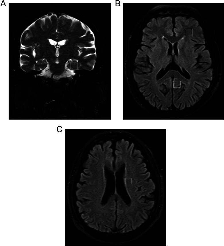
Figure 1.
Localized proton magnet resonance spectroscopy (MRS) of patient: (A) hippocampus (HIP); (B) white matter of the frontal lobe (FLWM) and posterior cingulate gyrus (PCG); (C) white matter adjacent to the lateral ventricles (WMALV). Region of interest (ROI) sizes were approximately 0.8 × 0.8 × 1.0 cm.

