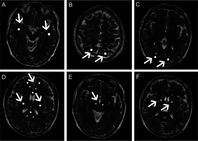
Figure 1.
Regions of interest (ROI) in white matter of patients with Parkinson’s disease. Apparent ROIs (the light regions indicated by white arrows) are shown in the (A) temporal white matter; (B) parietal white matter; (C) occipital white matter; (D) frontal white matter and anterior/posterior cingulate bundles, genu and splenium of the corpus callosum, and superior longitudinal fasciculus; (E) corticospinal tract in midbrain; (F) corticospinal tract in internal capsule of patients with Parkinson’s disease.

