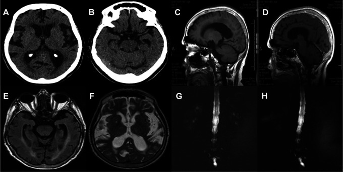
Figure 1.
A, A brain computed tomography (CT) revealed ventricular enlargement out of proportion to sulcal enlargement and cerebral atrophy with an Evan’s index of 0.31. Also, the image revealed periventricular signal changes as the evidence of altered brain water content in the patient who has no ischemic risk factors. B, The CT scan shows enlargement of the temporal horns of the lateral ventricles not entirely attributable to hippocampus atrophy. C and D, Sagittal views of T1-weighted and gadolinum-enhanced T1-weighted magnetic resonance imaging (MRI) revealed normal in terms of intracranial hypotension syndrome (IHS). E, Flair T2 axial image showed lateral ventricular enlargement and presence of an aqueductal flow void. F, T2-weighted axial image showed periventricular signal changes as in (A). G and H, Magnetic resonance myelography proved no evidence of cerebrospinal fluid leak.

