
Figure 2.
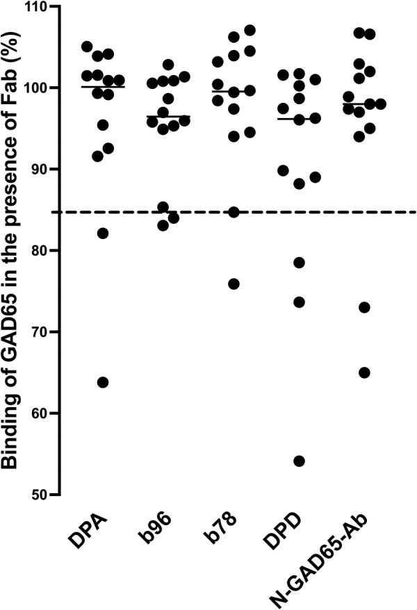
GAD65Ab epitope pattern in 14 GAD65Ab-positive T2D patients does not resemble that found in T1D patients or LADA patients. Binding of serum samples to GAD65 was evaluated in the presence of rFab DPA, b96.11, b78, DPD, and N-GAD65-Ab, and is reported as the percentage of uncompeted binding (set at 100%). The percentage bound remaining after competition with each rFab is presented for each sample. Short solid horizontal lines indicate median binding to each epitope. The cutoff value for successful competition is indicated by the dashed horizontal line. (Note: some dots directly overlie others in the DPD and N-GAD65-Ab columns.) The data indicate that the GAD65Ab epitope patterns in these GAD65Ab-positive T2D patients does not resemble those typically found in T1D or LADA patients.