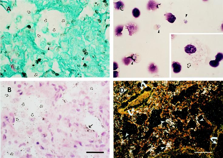
FIG. 1.
Pathology of P. carinii-infected lung tissue. Alveolar macrophages (open arrows) were detected in P. carinii-infected ferret lung tissue by GMS (A) and H&E (B) staining (cysts, arrowheads; trophs, arrows). Diff Quik staining demonstrates both trophic (inset, open arrow) and cyst (arrowheads) nuclei in a cytospin preparation of infected ferret lung homogenate (C). Note that the cyst forms of P. carinii shown in this field (arrowheads) contain between four and eight nuclei representative of intracystic bodies that correspond to the individual nuclei of the trophic forms released upon excystation during a stage in the P. carinii life cycle. The bar in panel B represents 20 μm for panels A to C. Trophic nuclei within a macrophage cytoplasm were occasionally detected (C, inset). (D) A low-power darkfield exposure of P. carinii-infected lung probed with the gpA antisense riboprobe specific for P. carinii gpA mRNA Bar, 70 μm. Arrows indicate blood vessels.

