
FIGURE 3.
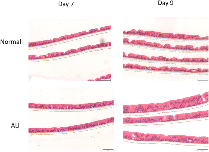
Micrographs of hematoxylin and eosin‐stained monolayers derived from different lines of human enteroids cultured in vitro under two different conditions, Normal(top), where the monolayer is submerged under the proliferation medium and ALI(bottom), where there is no medium on top of the monolayer. These are seen under bright field under 200× magnification. Scale bar: 50 μm.