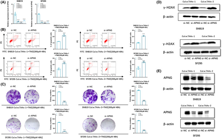
FIGURE 3.

Inhibiting APNG expression promoted TMZ‐induced cell apoptosis. (A) qPCR analysis of APNG expression in SNB19 and SF295 cells with si‐NC, si‐APNG‐1, si‐APNG‐2, si‐APNG‐3 transfection, respectively. (B) Flow cytometry analysis of SNB19 or SF295 cells after co‐culturing with TAAs followed with APNG knockdown and TMZ exposure (300 μM for SNB19, 200 μM for SF295) for 48 h. (C) Soft agar colony formation assay on SNB19 or SF295 cells after co‐culturing with TAAs followed with APNG knockdown and TMZ exposure (300 μM for SNB19, 200 μM for SF295) for 48 h. (D) Western blot of γ‐H2AX expression in SNB19 or SF295 cells after co‐culturing with TAAs followed with APNG knockdown and TMZ exposure (300 μM for SNB19, 200 μM for SF295) for 48 h. (E) APNG protein level of SNB19 or SF295 cells after co‐culturing with TAAs followed with APNG knockdown. **p < 0.01, ***p < 0.001, and ****p < 0.0001.
