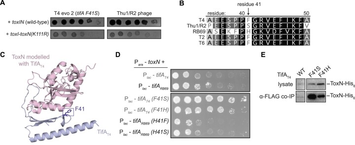
Figure 3.
Mutations in TifA that strengthen its interaction with ToxN. (A) Spotting assays for evolved T4 phage with increased expression of dmd-tifA(F41S) (T4 evo 2) and Thu1/R2 phage on lawns of +toxIN and +toxI-toxN(K111R) cells. (B) Alignment of residues 35–50 of TifA homologs from several T4-like phages, with residue 41 indicated. Degree of sequence similarity is highlighted in gray/black. (C) AlphaFold model for the ToxN-TifAT4 complex, with residue F41 on TifA highlighted. (D) Serial dilutions of E. coli cells ectopically expressing toxN and wild-type or mutant tifAT4 or tifARB69. (E) Western blot of ToxN-His6 in cell lysate and following co-immunoprecipitation with TifAT4-FLAG (wild-type or mutant) during T4 infection.

