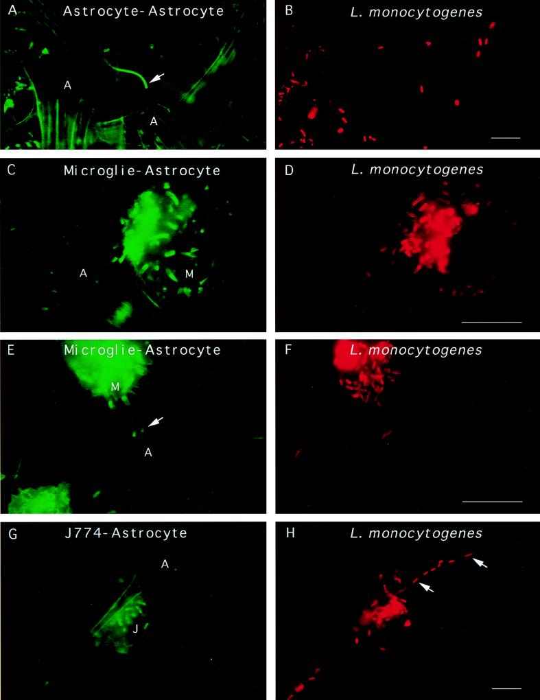
FIG. 3.
L. monocytogenes can move and spread from cell to cell in glial cell cultures. Glial cells were incubated with L. monocytogenes for 40 min, unbound bacteria were washed away, and the cells were resuspended in medium containing gentamicin and incubated for 19 h. The cells were fixed and stained for F-actin with FITC phalloidin. The bacteria were revealed by the technique of differential immunostaining to distinguish between extracellular (green and red) and intracellular (only red) bacteria. Panels A, C, and E show three different fields in which Listeria cells are clearly seen projecting away from astrocytes or microglial cells. Typical Listeria actin tails are indicated by arrows. Panels B, D, and F show the labeling of the bacteria in red. (G and H) J774 macrophages previously infected with L. monocytogenes were cultured with primary glial cell cultures for 19 h in a cell culture medium containing gentamicin to kill extracellular listeriae. The cells were fixed and stained for F-actin, and the presence of intracellular bacteria was determined by differential immunofluorescence. Panel G shows an infected macrophage near an astrocyte. Panel H shows the presence of intracellular L. monocytogenes in an astrocyte (thick arrows). Scale bar, 5 μm.

