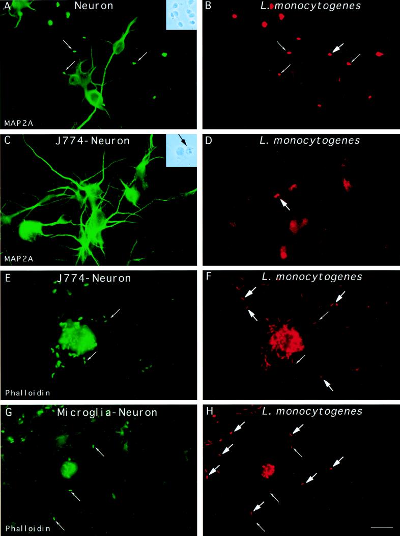
FIG. 4.
Entry of L. monocytogenes into cultured neurons. (A and B) Neurons were incubated with L. monocytogenes for 40 min, unbound bacteria were washed away, and the cells were resuspended in medium containing gentamicin and incubated for 2 h. The cells were fixed and stained with a MAb raised against MAP-2A (panel A) and against the bacterial pathogen. Extracellular bacteria are indicated by thin arrows and are both green and red, whereas intracellular bacteria indicated by thick arrows are only red. The unique intracellular bacterium that we found in neurons is shown in panel B. Insets: phase-contrast microscopy of primary cultures of neurons infected with L. monocytogenes (A) and of neuron-infected J774 macrophage cocultures (B). The black arrow shows a macrophage adjacent to a neuron. (C through F) J774 macrophages previously infected with L. monocytogenes were cultured with neurons for 19 h in tissue culture medium containing gentamicin. The cells were fixed and stained with a MAb raised against MAP-2A (panel C) or stained for F-actin (panel E), and the presence of intracellular bacteria was examined by differential immunofluorescence. Panel C shows neurons. Panel D shows the presence of two intracellular L. monocytogenes in a neuron. Panel E shows an infected macrophage and the actin network of the neurons. Panel F shows the presence of intracellular L. monocytogenes cells in neurons close to the infected macrophage. (G and H) Cultured neurons were infected with L. monocytogenes for 40 min, washed, and incubated for 15 h in L15 complete medium containing gentamicin. Cells were fixed and stained for F-actin, and the presence of intracellular bacteria was determined by differential immunofluorescence. Panel G shows a microglial cell in the neuronal monolayers. In the neuronal processes located in the vicinity of the infected microglial cell, intracellular bacteria, indicated by thick arrows in panel H, were detected. Scale bar, 10 μm.

