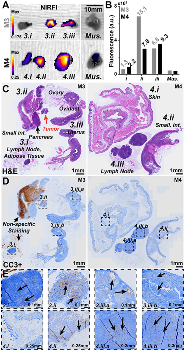
Figure 5. Necropsy and histological analysis of additional regions of interest during CT26 tumor resection.

A) During CT26 resection and necropsy small areas (labelled as 3.1, 3.2, etc.) were identified and found to be highly fluorescent (NIRFI, IVIS Spectrum, 745nm excitation, 840nm emission) over background areas and muscle tissue. B) Quantification of ROIs with ROI to muscle ratios shown (italics). C) Left, H&E staining of resected tissues from M3 identified (clockwise) as a lymph node surrounded by adipose tissue (3.1), the small intestine with pancreas and a small neoplastic tumor area not bound to any identifiable tissue (3.2) and finally the reproductive organs including the uterus, oviduct, ovaries (3.3). Right, H&E staining of resected tissues from M4 identified (clockwise) as the skin and subcutis (3.1), a lymph node surrounded by adipose tissue (3.2) and finally the small intestine also with adipose tissue (3.3). D) Cleaved caspase 3 positive (CC3+, for apoptosis i.e., damaged cells) IHC staining on a consecutive slice for each mouse sample. Labelled boxes highlight ROIs with increased levels of CC3 specific staining. Non-specific CC3 staining widely seen in the small intestine of M3 should be ignored. E) Zoomed in areas as highlighted at various magnification levels: 3.1 Small to moderate number of CC3+ cells were seen in the lymph node cortex and paracortex. 3.2 CC3+ cells were found in the tumor region. 3.3a CC3+ cells are highlighted in the granulosa of ovarian follicles. 3.3b Small number of CC3+ cells are seen within the endometrial epithelium. 4.1 No CC3+ cells were seen in the skin. 4.2 Moderate number of CC3+ cells are seen within the crypts with a small number in the enterocytes of villi. 4.3a & 4.3b Small to moderated number of CC3+ cells were seen throughout lymph node cortex and paracortex.
