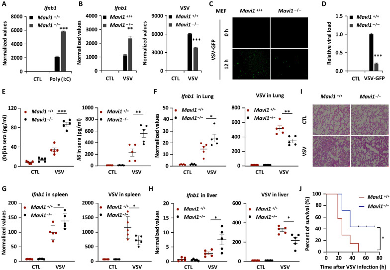
Fig. 3. MAVI1 negatively regulates RLR-mediated antiviral immune responses in vivo.
(A) WT (Mavi1+/+) and Mavi1 knockout (Mavi1−/−) MEFs treated with or without poly (I:C) (5 μg/ml, 6 hours) were subjected to examination of Ifnb1 expression (mean ± SEM, ***P < 0.001). (B) Mavi1+/+ and Mavi1−/− MEFs treated with or without VSV (2 × 10⁶ p.f.u., 12 hours) were subjected to examination of the expression of genes as indicated (mean ± SEM, **P < 0.01, ***P < 0.001). (C) Mavi1+/+ and Mavi1−/− MEFs were treated with VSV-GFP for 12 hours, followed by microscopy imaging analysis. (D) The number of GFP+ cells as shown in (J) were analyzed by ImageJ and normalized to Mavi1+/+ MEFs without VSV-GFP treatment. (E) The levels of Infβ and Il6 in serum from Mavi1 +/+ or Mavi1−/− mice (n = 5) treated with or without VSV (5 × 10⁸ p.f.u. per mouse) for 12 hours were examined by enzyme-linked immunosorbent assay (mean ± SEM, **P < 0.01, ***P < 0.001). (F to H) The levels of Infb and VSV from the lung (F), spleen (G), and liver (H) of mice as described in (L) were examined (mean ± SEM, *P < 0.05, **P < 0.01). (I) Lung tissue sections from mice as described in (M) were subjected to hematoxylin and eosin staining. (J) Survival curve of Mavi1 +/+ or Mavi1−/− mice (n = 7) treated with VSV (5 × 10⁸ p.f.u. per mouse) is shown (mean ± SEM, *P < 0.05).

