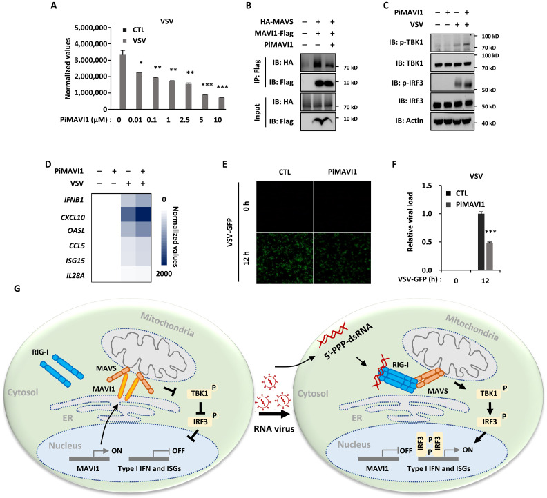
Fig. 8. Peptide inhibitor targeting the interaction between MAVI1 and MAVS (PiMAVI1) is potent in activating type I IFN signaling and antiviral immune responses.
(A) HEK293 cells were pretreated with peptide inhibitor PiMAVI1 at concentrations as indicated for 8 hours and then infected with or without VSV treatment (2 × 10⁶ p.f.u., 12 hours) were subjected to examination of VSV replication (mean ± SEM, *P < 0.05, **P < 0.01, ***P < 0.001). (B) HEK293 cells transfected with hemagglutinin (HA)–tagged MAVS and Flag-tagged MAVI1 and treated with or without PiMAVI1 (10 μM, 8 hours) were subjected to immunoprecipitation (IP) with anti-Flag antibody followed by immunoblotting (IB) analysis. (C and D) HEK293 cells pretreated with PiMAVI1 (10 μM, 8 hours) and then infected with VSV (12 hours) were subjected to IB (C) or RT-qPCR analysis (D). (E) HEK293 cells were pretreated with or without PiMAVI1 (10 μM, 8 hours) and then infected with or without VSV-GFP (2 × 10⁶ p.f.u., 12 hours), followed by microscopy imaging analysis. (F) The number of GFP-positive (GFP+) cells as shown in (E) was analyzed by ImageJ and normalized to control cells. (G) The proposed model of MAVI1 function in antiviral innate immune responses. ER-localized TM protein MAVI1 contacts with MAVS protein on mitochondria to compete with RIG-I for binding to MAVS, resulting in the repression of type I IFN signaling and innate immune responses. Upon VSV infection, MAVI1 is down-regulated, and its inhibition on the interaction between RIG-I and MAVS is released, leading to the activation of type I IFN signaling and innate immune responses. Peptide inhibitor targeting the interaction between MAVI1 and MAVS is capable of disrupting MAVI1-MAVS interaction, activating type I IFN signaling, and enhancing host antiviral innate immunity.

