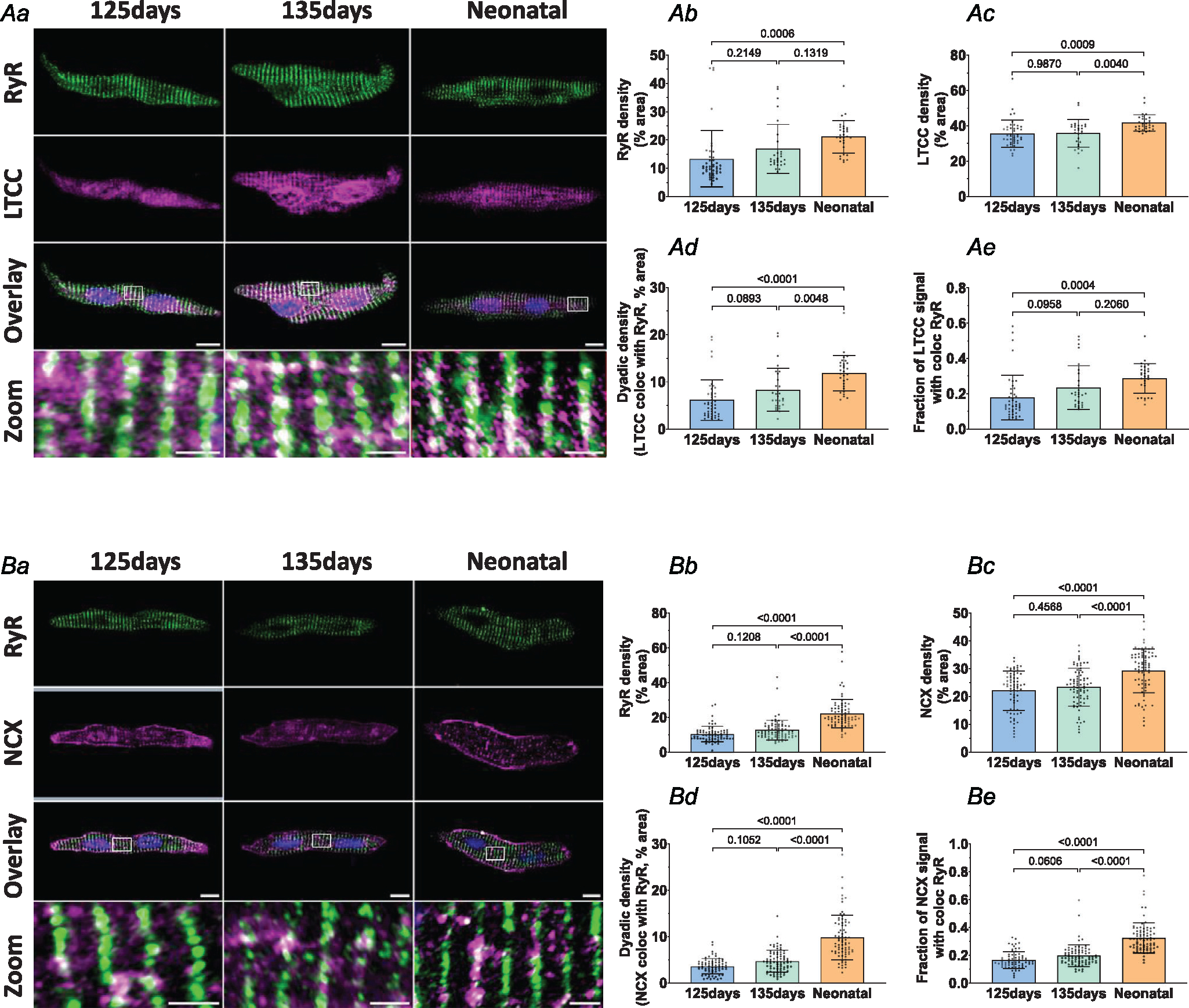
Figure 2. Cardiac dyads are progressively assembled during the fetal period.

Representative super-resolution Airyscan micrographs of isolated cardiomyocytes labelled with antibodies against RyR in combination with either LTCC or NCX (A and B, respectively, scale bars: 10 μm). DAPI was used to visualize the nuclei. For the indicated regions in the overlay images, enlargements are presented in the lower panels (scale bars: 2 μm). Image analysis revealed progressive increases in RyR density (Ab and Bb), LTCC density (Ac), and NCX density (Bc) in the developing heart. Gradual assembly of dyads was confirmed by increasing density of sites where either LTCC (Ad) or NCX (Bd) colocalized with RyRs. The proportion of LTCC (Ae) and NCX (Be) signals colocalized with RyRs showed a similar pattern of increase during development. Data are presented as mean ± SD, with P-values indicated. Statistical comparisons were made using 1-way ANOVA with Tukey’s multiple comparison test. Number of cells (animals): A: 125 days 43 (4), 135 days 29 (4), 1–5 days neonatal 30 (3); B: 125 days 69 (4), 135 days 77 (6), 1–5 days neonatal 77 (5).
