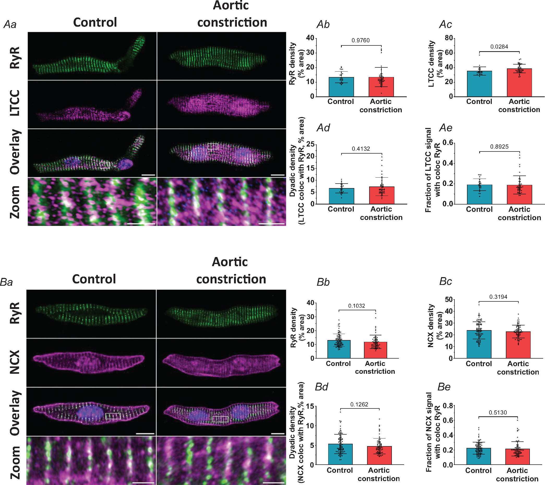
Figure 7. Cardiac dyads are not remodelled by increasing systolic load in the fetal heart.

Representative super resolution Airyscan micrographs of isolated cardiomyocytes labelled with antibodies against RyR in combination with either LTCC or NCX (A and B, respectively, scale bars: 10 μm). DAPI was used to visualize the nuclei. For the indicated regions in the overlay images, enlargements are presented in the lower panels (scale bars: 2 μm). Although t-tubule growth in the aortic constriction hearts was linked to increased LTCC density (Ac), no changes were detected in RyR density (Ab and Bb), NCX density (Bc), or dyadic density, as defined by the association of either LTCC (Ad) or NCX (Bd) with RyRs. The fraction of LTCC or NCX signal co-localized with RYR was also unchanged (Ae and Be). Data are presented as means ± SD, with P-values indicated. Statistical comparisons vs. control were made by unpaired t test. Number of cells (animals): A: controls 24 (3), aortic constriction 44 (4); B: controls 85 (6), aortic constriction 70 (5).
