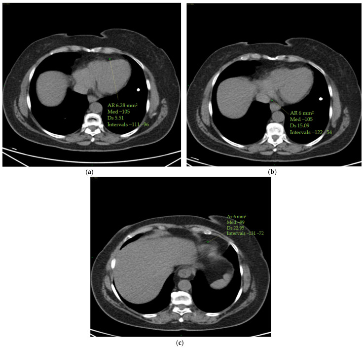
Figure 2.
(a–c) Epicardial fat density measurement. Using a 4-chamber projection, epicardial fat density was assessed on axial perspective basal CT scans, using a manually placed ROI (with a mean area of 6 mm2) within the anterior interventricular sulcus (a), posterior interventricular sulcus (origin of the posterior interventricular artery) (b) and cardiac apex (c). The figure shows an example of the area of the region of interest (Ar ROI), the mean (med), the standard deviation (Ds) and the range (intervals) of hounsfield units measured.

