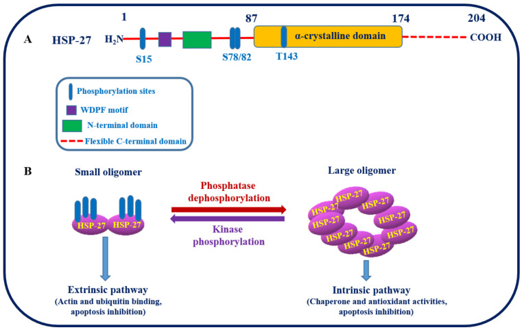
Figure 2.
(A) Structure of HSP27 and its putative phosphorylation sites. (B) Schematic representation showing the two different conformational states of HSP27 induced by phosphorylation, that is, large oligomers when unphosphorylated and small oligomers when phosphorylation is catalyzed by MAPKAPK 2/3 kinase at specific serine residues of HSP27. These conformational, structural, and functional changes actively contribute to the maintenance of cellular proteostasis [49].

