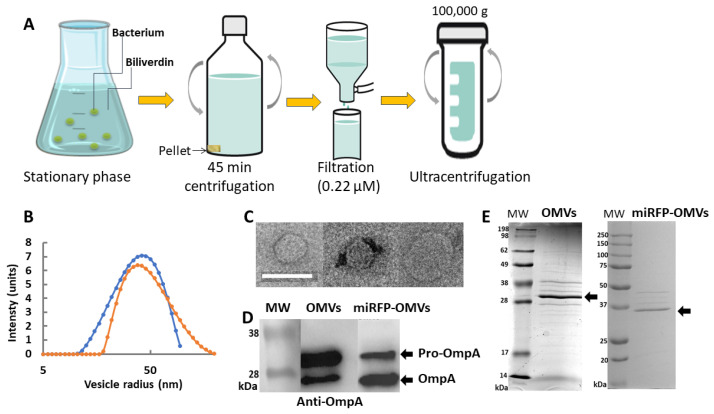
Figure 2.
In vitro production and characterization of OMVs secreted from E. coli REL606 versus E. coli miRFP713-expressing REL606. (A) Schematic representation of the various steps of production and purification of fluorescent miRFP713-OMVs versus non-fluorescent OMVs. (B) DLS distribution of OMVs (in blue) and miRFP713-OMVs (in orange) radii according to their frequencies. (C) Transmission electronic microscopic observations of 3 OMVs. Scale bar: 100 nm. (D) Immunoblot detection of the OmpA protein (~27 kDa for the mature protein, ~35 kDa for the pro-protein, black arrow heads) in OMVs (OMVs) versus miRFP713-OMVs (miRFP-OMVs). (E) Coomassie blue-stained SDS-PAGE gels of OMVs versus miRFP713-OMVs (miRFP-OMVs) proteins. The OmpA protein is indicated. MW: molecular weight.

