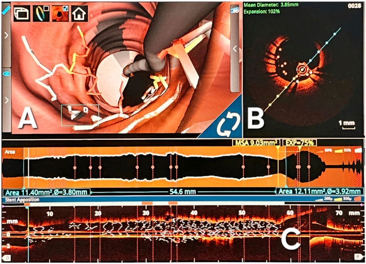
Figure 4.
OCT images used for optimal stent optimization. (A) Comprehensive three-dimensional intravascular reconstruction, vividly displaying instances of strut malposition; (B) Transversal section delineating the specifics of strut malposition, quantifying it at a length of 0.6 mm. This sectional view augments the understanding of the spatial irregularities within the stent deployment. (C) Longitudinal 3D reconstruction highlighting the stent along its entire length; significant malposition is observed at the distal end of the stent; D-Longitudinal reconstruction of the stent.

