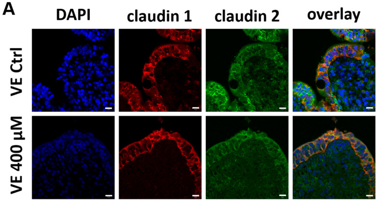
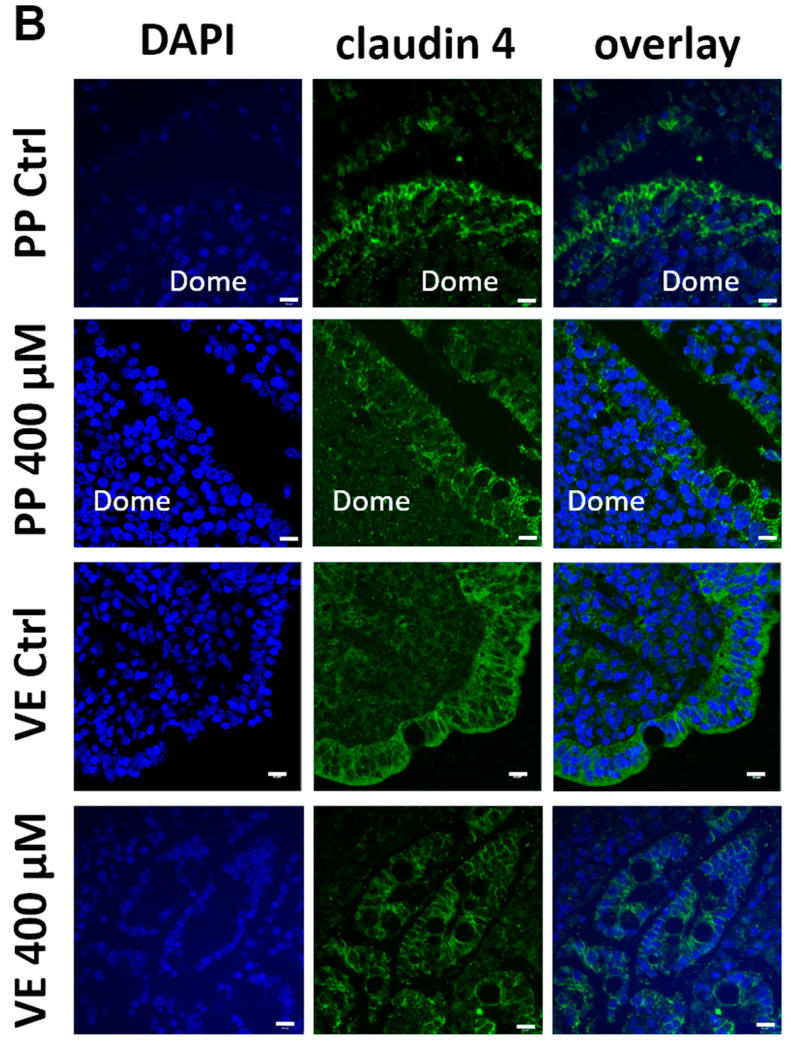
Figure 5.
Immunohistochemistry: Immunolocalization of tight junction proteins after 4 h incubation with or without 400 µM quercetin. (A) Confocal laser-scanning microscopy of claudin 1 (red) and claudin 2 (green) in the VE samples. (B) Widefield microscopy (PP Ctrl and VE 400 µM) and confocal laser-scanning microscopy (PPs 400 µM and VE Ctrl) of claudin 4 (green) in PPs and VE samples. The tight junction proteins can be detected as paracellular signals in the surface epithelium. Cell nuclei were stained blue with DAPI (scale bar: 10 µm, n = 5, representative images).


